Fully Guided Implant Surgery
There are a lot of dentists placing dental implants these days, and there are several different methods of placing them. I’d like to share the basics of these methods and hopefully enable those of you who are considering dental implants to better understand what is involved in the their placement. Next month, I’ll share some concepts associated with the successful restoration (affixing the teeth) of implants.
In the beginning, implants were placed free hand and wherever bone was located. The challenge with planning dental implant position based on available bone is that available bone does not always allow for an ideal crown position. A crown placed on a poorly positioned implant can appear misshapen, out of place, and have damaging force placed on it due to its unbalanced positioning. The current thinking is to establish the ideal tooth position first, then evaluate if there is enough available bone to place the implant in a way that will allow that ideal tooth position. If there is enough bone available, all is well. However, if there is inadequate bone in the area, some amount of bone grafting must be done in order to allow an implant placement that will support the proper final tooth position.
In spite of many advances in treatment planning and surgical precision, many implants are still placed free hand. One reason for this may be the relative simplicity of a given situation. Consider, for example, the single missing molar or premolar with adjacent remaining teeth and plentiful bone in which to place the implant. For an experienced implantologist, this is a relatively simple case and not much will be gained by adding the additional expense of a surgical guide. That being said, a properly planned surgical guide will reduce surgical stress, surgical time, and will greatly improve chances of successful outcomes.
A surgical guide used to be made from a stone model of the patient’s mouth. The laboratory would use the model to plan out the proper tooth position and make a pilot hole guide that would allow the implantologist the ability to snap the guide on the patient’s teeth and know where to make the initial hole. Two big challenges with this style of guide were that there was little consideration as to whether there was enough bone in the area and they only allowed guidance for the initial drill hole into the bone. At the time, this was good. Thankfully, improved imaging technology has improved both the ability to evaluate available bone and to make a very precise surgical guide based on that evaluation.
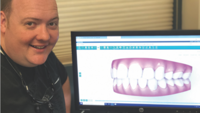
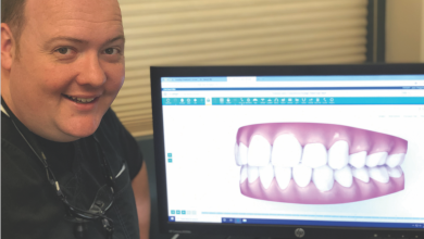
2-D imaging has been used for years to evaluate dental implant sites. In recent years, with the advent of cone beam computed tomography (CBCT), 3-D evaluation has become a reality. This allows for precise understanding of nerve positioning, sinuses, undercuts in bony contour, and more. As the technology has continued to develop, the ability to virtually place an implant in a patient on a computer monitor has enabled us to make a surgical guide that allows for the placement of the implant in that precise, pre-surgically planned position.
The current state-of-the-art is “fully guided” implant placement. Many guides require removal at some stage in order to place the final implant. A fully guided approach plans the implant position with the CBCT image virtually, fabricates a guide based on the positioning, and allows the implantologist to place the guide in the patient’s mouth and go from initial hole to final implant placement – controlling the position, angulation, and depth of that placement.
I have placed dental implants free hand, with stone model surgical guides, with CBCT surgical guides, and with CBCT fully guided techniques. And while I feel very confident in the free hand placement of dental implants in the right circumstances, I am very excited about the future of fully guided implant placement and what it will enable implantologists to provide for patients in the rehabilitation of their oral health and function.
I hope this has been helpful :^)


